Sacroiliac Joint
The SI joint is the articulation between the ilium and the sacrum on each side of the pelvis. As with other joints, it is comprised of the bony stabilizers, the static soft tissue or ligamentous stabilizers, and the dynamic muscular stabilizers. On the surface of the bone is the articular cartilage.
The SI joint depends primarily on the stout ligaments that cross it for stability. The bones also have shallow interdigitations that correspond on each side, thus conferring some bony stability. Finally, there are the muscles (dynamic stability) and fascia—especially the thoracolumbar fascia.
Figure 1 illustrates the bones that comprise the SI joint.
Figure 2 illustrates the stout ligamentous stabilizers of the joint. These include:
Movement is very limited for this joint, but includes nutation or anterior tilt (flexion) of the sacrum between the ilia, counter-nutation or posterior tilt (extension) and small movements of the ilia themselves. The stable SI joint thus functions for shock absorption and transfer of torque during ambulation.
Muscles and fascia also confer stability to the joint. Figure 3 illustrates the relationship between the erector spinae muscles of the back and the muscles of the pelvic floor. You can see that the erector spinae muscles draw the sacrum into flexion (nutation) and the muscles of the pelvic floor (especially the pubococcygeus) draw the bone into extension (counter-nutation). Simultaneously engaging these muscles creates opposing forces that stabilize the joint.
Figure 4 illustrates the relationship of the latissimus dorsi and gluteus maximus muscles on opposite sides of the body. In between is the thoracolumbar fascia. Note how the fibers of these structures run perpendicular to the joint. Thus, working with core exercises such as Bird Dog Pose can help to strengthen the dynamic stabilizers of the SI joint. These muscles, along with the fascia comprise the “posterior oblique subsystem”.
The SI joint depends primarily on the stout ligaments that cross it for stability. The bones also have shallow interdigitations that correspond on each side, thus conferring some bony stability. Finally, there are the muscles (dynamic stability) and fascia—especially the thoracolumbar fascia.
Figure 1 illustrates the bones that comprise the SI joint.
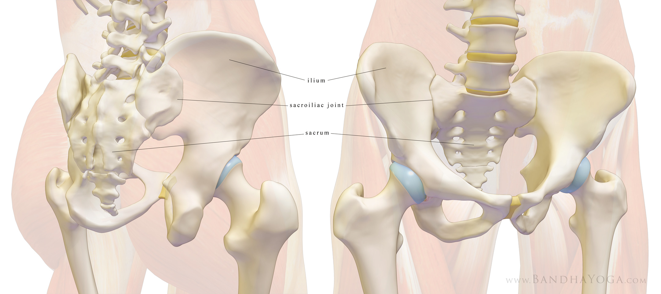 |
| Figure 1: The bones of the sacroiliac joint. |
Figure 2 illustrates the stout ligamentous stabilizers of the joint. These include:
- The anterior (front) and posterior (back) sacroiliac ligaments running from the sacrum to the ilium;
- The sacrotuberous ligaments running from the sacrum to the ischial tuberosity;
- The sacrospinous ligaments running from the sacrum to the posterior iliac spine;
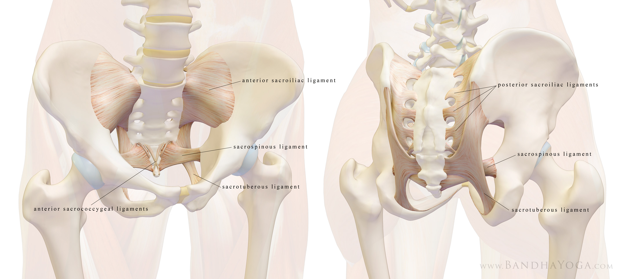 |
| Figure 2: The ligaments of the sacroiliac joint. |
Movement is very limited for this joint, but includes nutation or anterior tilt (flexion) of the sacrum between the ilia, counter-nutation or posterior tilt (extension) and small movements of the ilia themselves. The stable SI joint thus functions for shock absorption and transfer of torque during ambulation.
Muscles and fascia also confer stability to the joint. Figure 3 illustrates the relationship between the erector spinae muscles of the back and the muscles of the pelvic floor. You can see that the erector spinae muscles draw the sacrum into flexion (nutation) and the muscles of the pelvic floor (especially the pubococcygeus) draw the bone into extension (counter-nutation). Simultaneously engaging these muscles creates opposing forces that stabilize the joint.
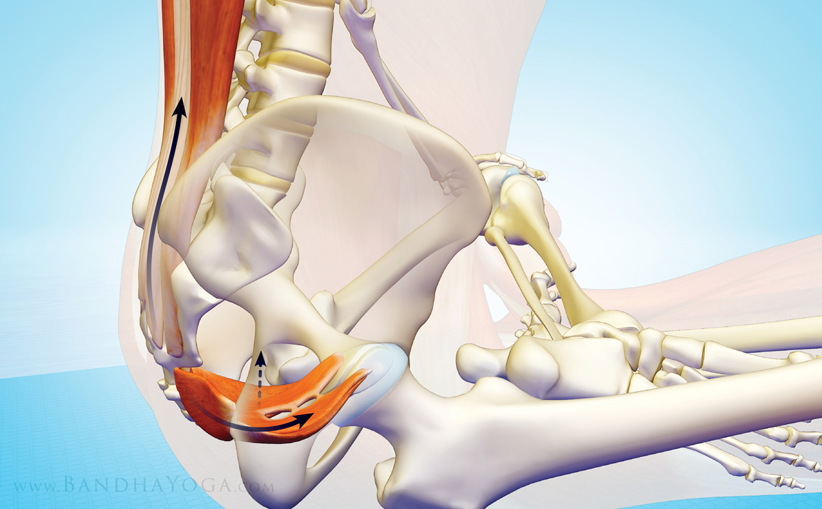 |
| Figure 3: The interaction between the erector spinae and pelvic floor muscles for stabilizing the SI joint. |
Figure 4 illustrates the relationship of the latissimus dorsi and gluteus maximus muscles on opposite sides of the body. In between is the thoracolumbar fascia. Note how the fibers of these structures run perpendicular to the joint. Thus, working with core exercises such as Bird Dog Pose can help to strengthen the dynamic stabilizers of the SI joint. These muscles, along with the fascia comprise the “posterior oblique subsystem”.
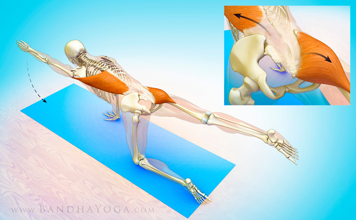 |
| Figure 4: The posterior oblique subsystem for stabilizing the SI joint. |